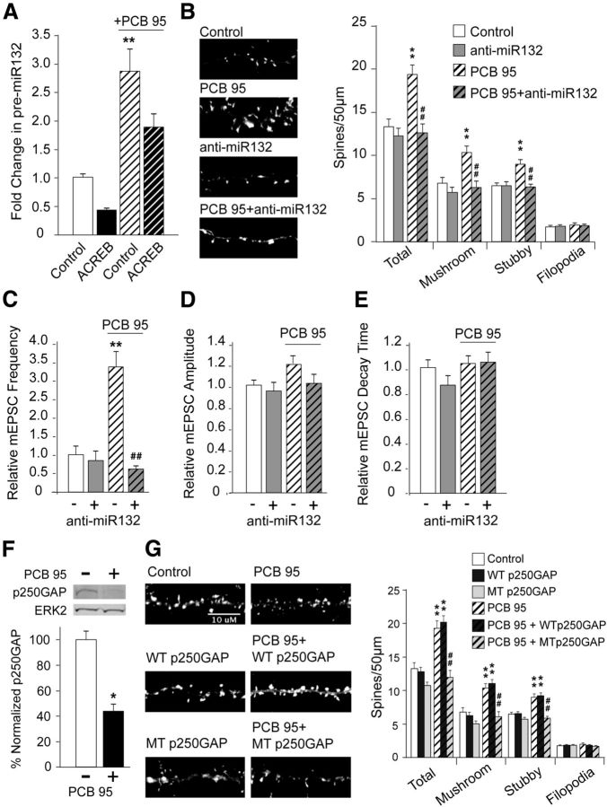
Figure 4.
miR132 is required for PCB 95-induced synaptogenesis. A, Dissociated hippocampal neurons were electroporated with empty expression vector or ACREB before plating and then cultured until DIV6 when they were treated for 2 h with vehicle or PCB 95 (200 nm). Pre-miR132 (normalized to HPRT) was determined using qPCR (n = 19–21). B–F, DIV6 dissociated hippocampal neurons transfected with mRFP-tagged β-actin ± empty vector or anti-miR132 were treated with vehicle or PCB 95 (200 nm) on DIV7-DIV12. B, Representative photomicrographs of RFP+ cells and quantification of dendritic spines and filopodia (n = 21–24 neurons). C–E, Quantitative analyses (n = 11–16 neurons) demonstrating PCB 95 effects on mEPSC frequency (C), amplitude (D), and decay time (E) represented as fold change relative to control cultures. F, Representative Western blot illustrating p250GAP protein levels in DIV6 dissociated hippocampal neurons treated with vehicle or PCB 95 (200 nm) for 24 h. Densitometric values of bands immunoreactive for p250GAP normalized to ERK2 levels within the same sample presented as percentage of control (n = 3 independent samples). G, Dissociated hippocampal neurons transfected on DIV6 with mRFP-tagged β-actin ± empty vector, WT p250GAP, or MT p250GAP were treated with vehicle or PCB 95 (200 nm) on DIV7–12. Representative photomicrographs of RFP+ cells and quantification of dendritic spines and filopodia (n = 23–24 neurons). *p < 0.05, significantly different from control. **p < 0.001, significantly different from control. ##p < 0.001, significantly different from PCB 95 alone (one-way ANOVA with post hoc Tukey's analysis).

