
Figure 5.
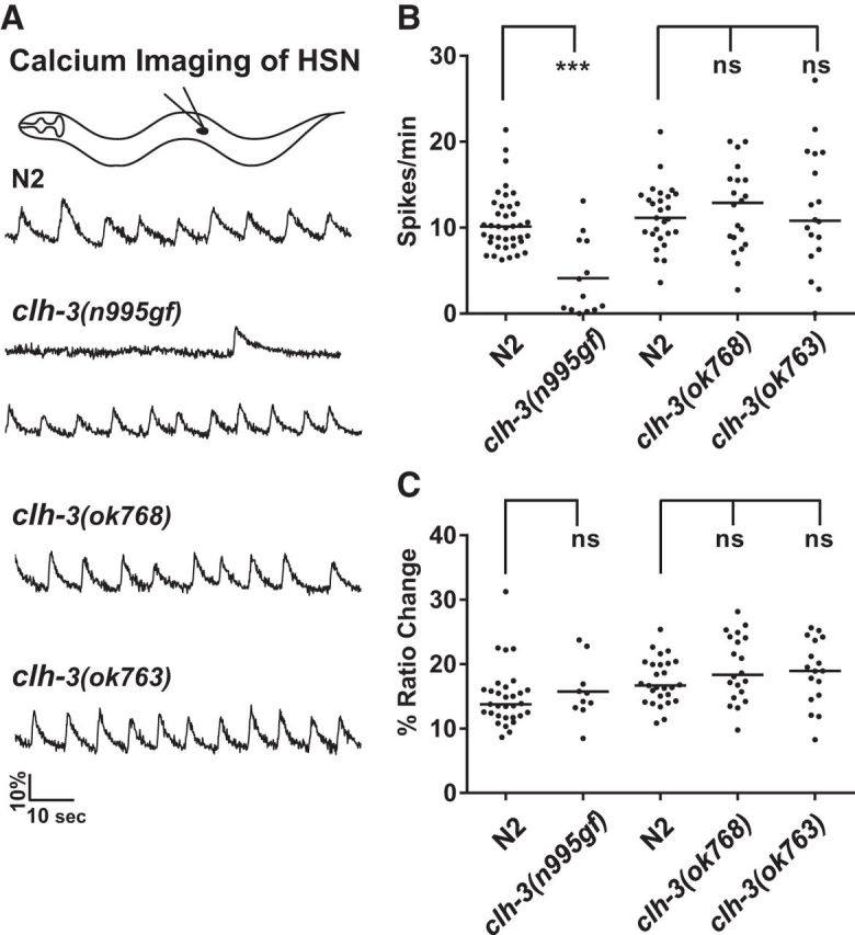
Calcium imaging reveals that CLH-3 inhibits HSN activity. A, Sample traces. The HSNs exhibit spontaneous rhythmic activity (Zhang et al., 2008). Most clh-3(n995gf) mutants have greatly reduced HSN activity, whereas some retain wild-type levels. B, C, Quantification of calcium spike parameters. The average number of spikes/minute and average percentage ratio change of each spike was determined for each recording. Each point represents the value obtained from one recording obtained from one animal. n = > 13 recordings, which corresponds to 145 spikes for clh-3(n995) and > 750 spikes for the other genotypes. Mutants were compared with the wild-type assayed at the same time. The clh-3(n995gf) mutation significantly reduces the spike frequency. None of the mutations alter spike parameters such as ratio change (as shown), area of spike, rise time, or decay time (data not shown).