Figure 1.
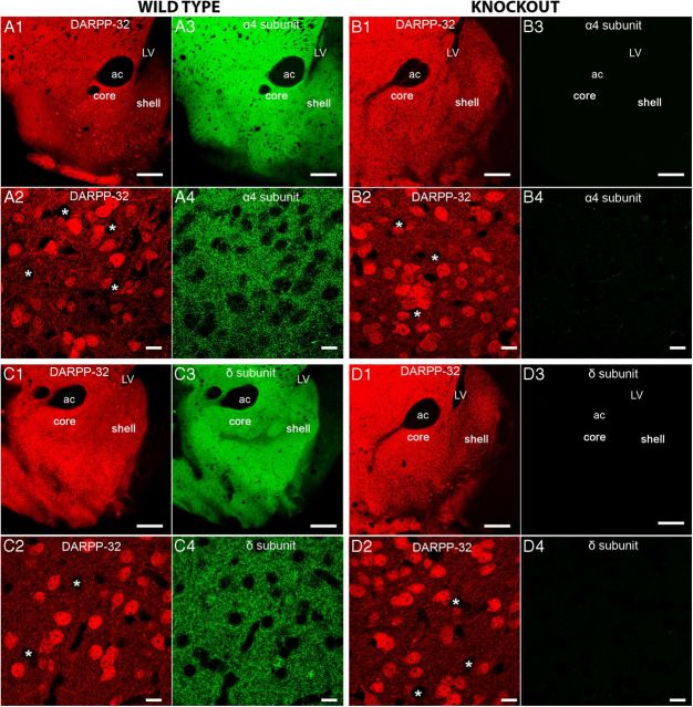
The distribution of GABAAR α4 and δ subunit immunoreactivity in the ventral striatum closely overlaps with the distribution of medium spiny neurons (MSNs). A, B, Representative images of immunoreactivity for the GABAAR α4 subunit and DARPP-32 in the ventral striatum, including the core and shell regions of the nucleus accumbens (NAc) in tissue from wild type (WT) and α4−/− mice, respectively. A1 shows a regional overview and A2 a magnified view of individual neurons expressing DARPP-32 immunoreactivity. DARPP-32 is only expressed in neurons that express dopamine receptors. Therefore, DARPP-32 immunoreactivity within the NAc is restricted to MSNs and not interneurons, which are likely to be those immunonegative neurons highlighted by the asterisks. A3 and A4 show the corresponding pattern of immunoreactivity for the GABAAR α4 subunit in the regions represented in A1 and A2. The GABAAR α4 subunit signal closely correlates with that of DARPP-32 in the NAc, suggesting the widespread expression of the α4 subunit throughout the MSNs of this brain area. Furthermore, there is no discernible variation in the expression of the α4 subunit throughout the NAc subregions. B1, B2, There were no detectable differences in the immunoreactivity pattern of DARPP-32 in tissue from WT and α4−/− mouse, respectively. B3, B4, No specific GABAAR α4 subunit signal was detected in tissue from GABAAR α4−/− mice. C1 shows a regional overview and C2 a magnified view of individual neurons expressing DARPP-32 immunoreactivity. C3 and C4 show the corresponding pattern of immunoreactivity for the GABAAR δ subunit in regions represented in C1 and C2. Immunoreactivity for the GABAAR δ subunit closely correlates with that of DARPP-32 in the NAc, suggesting widespread expression of the δ subunit throughout the MSNs of this brain area. Similar to the α4 subunit, there is no discernible variation in the expression of the δ subunit throughout the NAc. D1, D2, There were no detectable differences in the immunoreactivity pattern of DARPP-32 in tissue from WT and GABAAR δ−/− mice. D3, D4, No specific GABAAR δ subunit signal was detected in tissue from GABAAR δ−/− mice. Scale bars: A1, A3, B1, B3, C1, C3, D1, D3, 300 μm; A2, A4, B2, B4, C2, C4, D2, D4, 10 μm. ac, Anterior commissure; LV, Lateral ventricle.

