
Figure 3.
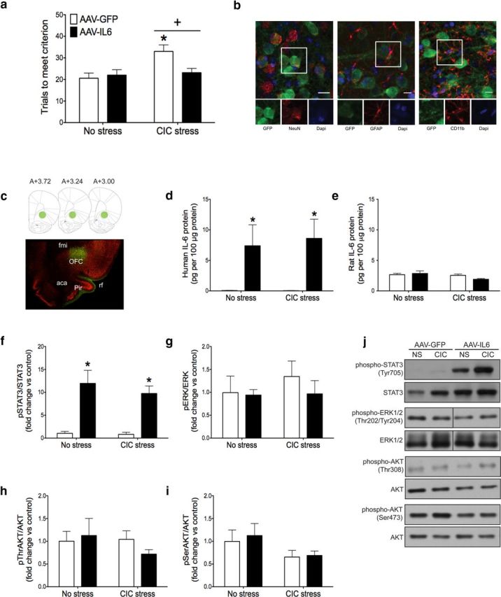
Effect of chronic IL-6 overexpression on reversal learning and downstream signaling pathways in the OFC. a, CIC stress increased trials to meet criterion on the reversal learning task, and AAV-IL-6 injection into the OFC restored reversal learning performance to nonstress levels. +p < 0.05, main effect compared with no stress control groups. *p < 0.05, compared with the AAV-IL-6-injected animals subjected to CIC stress. b, AAV-GFP exclusively infected neurons, determined by GFP expression seen in neurons but not astrocytes or microglia. Scale bar, 10 μm. c, Representative micrograph showing the extent of infection in the OFC, with corresponding diagrams at AP levels from bregma 3.72 to 3.00 (Paxinos and Watson, 2006). Green represents GFP; red represents NeuN. d, e, AAV-IL-6 increased the expression of recombinant human IL-6 in the OFC, without affecting endogenous rat IL-6 levels. *p < 0.05, main effect compared with AAV-GFP-infected animals. f–i, AAV-IL-6 injection increased STAT3 phosphorylation in the OFC without affecting ERK1/2 or AKT phosphorylation. Fold change relative to No Stress, AAV-GFP control group. *p < 0.05, main effect compared with AAV-GFP-injected animals. j, Representative Western blot images. fmi, Forceps minor of the corpus callosum; rf, rhinal fissure; aca, anterior commissure; Pir, piriform cortex. n = 7 or 8 per group.