
Figure 3.
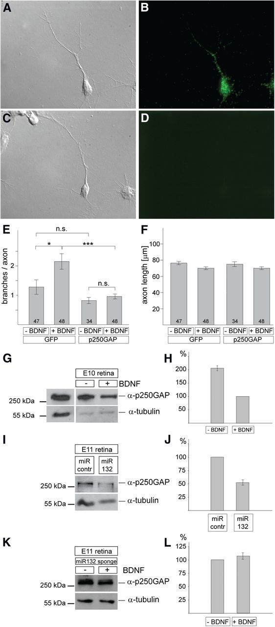
Analysis of p250GAP in chick retinal cultures. A, B, E10 chick RGC axons were stained with an anti-p250GAP antibody (B), and the corresponding differential interference contrast (DIC) picture is shown in A. C, D, A control staining omitting the anti-p250GAP antibody is shown in D with the corresponding DIC picture in C. E, Quantification of the number of branches of RGC axons after expression of p250GAP or GFP in the presence or absence of BDNF. The total number of axons analyzed for each condition is given. Overexpression of p250GAP abolishes any increase in BDNF-mediated branching. F, There is no statistically significant difference in the axon length under the various conditions. E, F, Error bars denote SEM. Significance is indicated as ***p < 0.001, *p < 0.05. n.s., Not significant. Statistical tests were performed with both Student's t test and Mann–Whitney. G, Single-cell preparations from chick E8 retina were incubated for 2 d in the presence or absence of 5 ng/ml BDNF, lysed, and analyzed for p250GAP expression by Western blot. For normalization, lysates were analyzed with an anti-α-tubulin antibody. Quantification is shown in H. The left lane shows an analysis of lysates from CHO cells transfected with the p250GAP expression plasmid to demonstrate the specificity of the antibody and the expected size of p250GAP. H, p250GAP expression is approximately twofold higher in the absence versus the presence of BDNF (average ± SEM, 2.06 ± 0.1) based on densitometric quantification of Western blots from three experiments. I, J, Single-cell preparations from E8 chick retinae were transfected with miRNA (miR)-132 or control miRNA expression plasmids and incubated for 3 d without adding BDNF. Lysates were subjected to Western blot analysis using anti-p250GAP antibodies and as a loading control anti-α-tubulin. P250GAP expression is reduced by miRNA-132 expression to 52.9 ± 9.3% relative to control miRNA expression (100%) based on densitometric quantification of Western blots from three experiments. K, L, Single-cell preparations from E8 chick retinae were transfected with miRNA-132 sponge plasmids (Fig. 2D–F) and incubated for 3 d with or without adding BDNF. Lysates were subjected to Western blot analysis using anti-p250GAP antibodies and as a loading control anti-α-tubulin. There was no statistically significant difference in p250GAP expression in the presence of BDNF (107.8 ± 7.4 relative to the expression of p250GAP in the absence of BDNF (100%) based on densitometric quantification of Western blots from three experiments. contr., Control.