Figure 4.

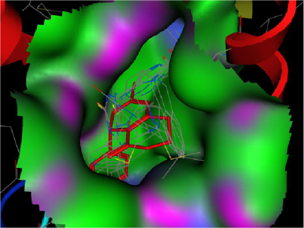
Map surface of docked analogs in active site of enzyme (Green: hydrophobic; Violet: H bonding; Blue: mild polar). Crystallized enasteron is indicated by red and stick lines.

Map surface of docked analogs in active site of enzyme (Green: hydrophobic; Violet: H bonding; Blue: mild polar). Crystallized enasteron is indicated by red and stick lines.