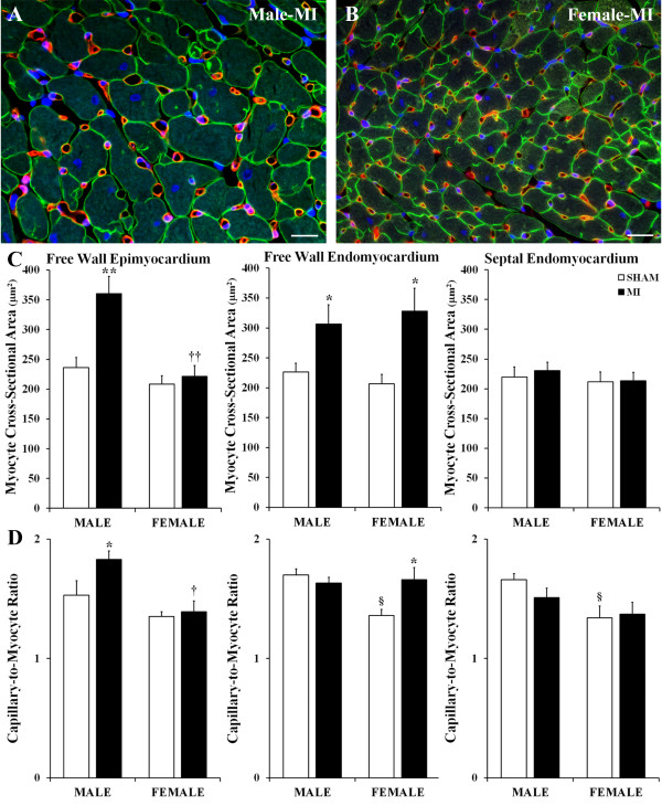
Figure 3.
Degree of reactive cardiac myocyte hypertrophy and compensatory angiogenesis. (A,B) Representative images of the free wall epimyocardium of post-MI male and female hearts immunofluorescence-stained with an antibody against laminin (green color), BSI isolectin B4 (red color), and DAPI (blue). (C) Cross-sectional area of cardiac myocytes in the LV free wall and septum. (D) Capillary-to-myocyte ratios in the LV free wall and septum. Scale bars are 20 μm. Values are the mean ± SEM; n = 7 male rats/group; n = 6 female rats/group. Note, in septal myocardium, all measurements were done on the endomyocardial side that is faced toward the LV cavity. A two-way ANOVA revealed a statistically significant interaction between the effects of sex and the experimental model on myocyte CSA in epimyocardial region, F (1, 22) = 6.032, P = 0.026, and on the capillary-to-cardiac myocyte ratio in free wall endomyocardium, F (1, 22) = 6.307, P = 0.025. *P < 0.05 and **P < 0.01 vs. a corresponding sham group; §P < 0.05 vs. M-Sham; †P < 0.05 vs. M-MI rats.

