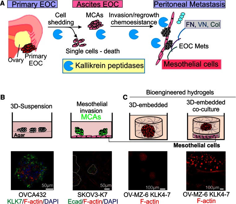
Fig. 2.
Cell culture platforms to mimic EOC metastasis. a Schematic diagram showing potential roles of KLKs in EOC peritoneal dissemination. b Top left panel, 3D-suspension cultures to mimic the EOC cells growing in the ascites fluid microenvironment. Top right panel, MCAs invading into a monolayer of mesothelial cells to mimic peritoneal invasion. Bottom left panel, MCAs formed by OVCA432 cells stained with anti-KLK7 and AlexaFluor488 (green), F-actin stained with AlexaFluor568 phalloidin (red), nuclei stained with 4′,6-diamidino-2-phenylindole (DAPI, blue). Bottom right panel, invasion into mesothelial cell monolayer by KLK7-expressing SKOV3 cell (SKOV3-K7) MCAs stained with anti-E-Cadherin (Ecad) and AlexaFluor488 (green), F-actin stained with AlexaFluor568 phalloidin (red), nuclei stained with DAPI (blue). c Top panel, schematic showing ovarian cancer cells embedded and cultured in 3D bioengineered hydrogels, alone (left) and sheet (right) co-culture with mesothelial cells. Bottom panel: KLK4–7-expressing OV-MZ-6 cells grown in the above models. F-actin filaments stained with rhodamine-415 conjugated phalloidin (red) and imaged by confocal laser scanning microscopy. Scale bars as indicated. With permission from Walter de Gruyter to use the elements of the figure in the published book ‘Kallikrein-related peptidases’ [114]

