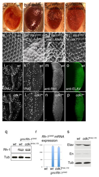
Figure 1. CDK7 and MAT1 mediate the mutant Rh-1G69D overexpression phenotype.
External eyes imaged by light microscopy (a–d) or Scanning EM (e–h). Wild type adult fly eyes show regular array of ommatidia (a, e), which is destroyed when Rh-1G69D is overexpressed during eye development through the eye-specific GMR promoter (b, f). In vivo RNAi screen for suppressors of this phenotype identified lines that target mat1 (c, g) and cdk7 (d). GMR-Rh-1G69D –induced ommatidial disruption phenotype was also suppressed in a cdk7S164A,T170A background (h). (i–l) eye imaginal discs were labeled for BrdU incorporation (i, j), anti-phospho Histone H3 antibody (k, l). (m,-p) GMR>Rh-1G69D eye discs were labeled with anti-Rh-1 antibody (m, n) and anti-ELAV antibody (o, p). These signals appear similar between cdk7 wild type (i, k, m, o) and cdk7S164A,T170A discs (j, l, n, p). (q) Western blots for Rh-1 and Beta-Tubulin (Tub) from wild type (lane 1) and GMR>Rh-1G69D (lanes 2 and 3) eye imaginal disc extracts. Lane 3 shows overexpressed Rh-1G69D levels in the cdk7164, 170background. (r) Quantitative RT-PCR against Rh-1G69D transcripts in genotypes equivalent to that of (q). (s) Western blots for ELAV, Engrailed (En) and Beta-Tubulin (Tub) in wild type and cdk7S164A,T170A imaginal disc extracts.

