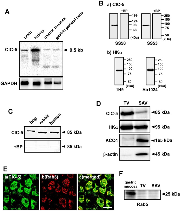
Fig. 1. Expression of ClC-5 in gastric samples.

(A) Expression of ClC-5 mRNA in the stomach. Northern blotting was performed with poly A+ RNA (2.5 µg/lane) from brain, kidney and stomach of rabbits. A single band of 9.5 kb was detected with the ClC-5 cDNA probe. As a control, expression of GAPDH (1.3 kb) was examined. (B) Specificity of anti-ClC-5 and anti-H+,K+-ATPase α-subunit (HKα) antibodies. Western blotting was performed with hog gastric tubulovesicles (10 µg of protein) by using anti-ClC-5 antibodies (SS58 and SS53) (a) and anti-HKα antibodies (1H9 and Ab1024) (b). A single band of 85 kDa (a) or 95 kDa (b) was detected. The 85-kDa band was disappeared when the ClC-5 antibodies were preincubated with the corresponding blocking peptide (antibody:peptide = 1:5) (+BP; a). (C) Western blotting was performed with hog and human gastric tubulovesicles and rabbit gastric P3 fraction (5, 50 and 40 µg of protein, respectively) by using anti-ClC-5 antibody (SS58). In each sample, an 85 kDa-band was detected (upper panel), and the band disappeared in the presence of the corresponding blocking peptide (lower panel). (D) Western blotting was performed with hog tubulovesicles (TV) and stimulation-associated vesicles (SAV) (10 µg of protein) by using anti-ClC-5 (SS58), anti-HKα (1H9), anti-KCC4 and anti-β-actin antibodies. ClC-5 (85 kDa) was predominantly expressed in TV, while KCC4 (165 kDa) and β-actin (45 kDa) were predominantly in SAV. HKα (95 kDa) was found in both TV and SAV. (E) a–c show the same tissue under a microscope. Double immunostaining was performed with hog gastric mucosa by using anti-ClC-5 (SS53) plus anti-Rab5 antibodies. Original magnification: ×63. Scale bars, 20 µm. (F) Western blotting was performed with TV, SAV and membrane fraction of gastric mucosa of hogs (30 µg of protein) by using anti-Rab5 antibody. Rab5 (25 kDa) was expressed in the gastric mucosa.
