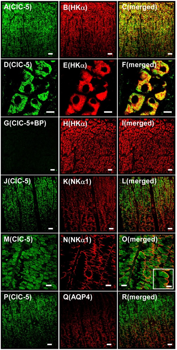
Fig. 2. Immunostaining for ClC-5 in isolated hog gastric mucosa.

A–C show the same tissue under a microscope (as do D–F, G–I, J–L, M–O and P–R). Double immunostaining was performed with hog gastric mucosa by using anti-ClC-5 (SS53) plus anti-HKα (1H9) antibodies (A–I), anti-ClC-5 plus anti-NKα1 antibodies (J–O), and anti-ClC-5 plus anti-AQP4 antibodies (P–R). (A–F) Localizations of ClC-5 (A and D), HKα (B and E) and ClC-5 plus HKα (merged image; C and F) are shown. (G–I) Anti-ClC-5 antibody was pretreated with the blocking peptide. Localizations of ClC-5 (G), HKα (H) and ClC-5 plus HKα (merged image; I) were shown. Positive ClC-5 staining disappeared (G). (J–O) Localizations of ClC-5 (J and M), NKα1 (K and N) and ClC-5 plus NKα1 (merged image; L and O). In the inset of O, an enlarged image of a parietal cell is shown. (P–R) Localizations of ClC-5 (P), AQP4 (Q) and ClC-5 plus AQP4 (merged image; R). Original magnification: ×20 (A–C, G–L and P–R), ×40 (M–O) and ×63 (D–F). Scale bars, 50 µm (A–C, G–L and P–R), 10 µm (D–F and inset of O), and 20 µm (M–O).
