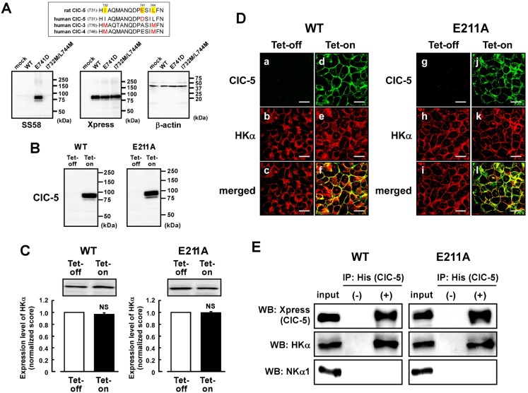
Fig. 4. Tetracycline-regulated expression of ClC-5 in the HEK293 cells stably expressing gastric H+,K+-ATPase.
(A) Alignments of rat ClC-5, human ClC-5, human ClC-3 and human ClC-4 around an epitope of the anti-ClC-5 antibody are shown (upper panel). WT-ClC-5, E741D-ClC-5 and I732M/L744M-ClC-5 were transiently transfected in the HEK293 cells. In lower panels, Western blotting was performed with the membrane fraction (50 µg of protein) using anti-ClC-5 (SS58) (left), anti-Xpress (middle) and anti-β-actin (right) antibodies. No significant signal was observed in mock-transfected cells. (B) The tetracycline-regulated expression systems of WT-ClC-5 and E211A-ClC-5 were introduced to the HEK293 cells stably expressing H+,K+-ATPase. The cells were treated with (Tet-on) or without (Tet-off) 2 µg/ml tetracycline. Expression of WT- and E211A-ClC-5 in the membrane fraction of the cells (30 µg of protein) was confirmed by Western blotting using anti-Xpress antibody. (C) Expression level of HKα in the Tet-on cells was compared with that in the Tet-off cells. In the upper panel, a representative picture of Western blotting is shown. In the lower panel, the quantified score for the Tet-off cells is normalized as 1. n = 6. NS, P>0.05. (D) a–c show the same cells under a microscope (as do d–f, g–i, j–l). Double immunostaining was performed with the WT Tet-off cells (a–c), WT Tet-on cells (d–f), E211A Tet-off cells (g–i) and E211A Tet-on cells (j–l) using anti-Xpress (for ClC-5) plus anti-HKα (Ab1024) antibodies. Localizations of WT-ClC-5 (a and d), E211A-ClC-5 (g and j) and HKα (b, e, h and k), WT-ClC-5 plus HKα (merged images; c and f), and E211A-ClC-5 plus HKα (merged images; i and l) are shown. Scale bars, 20 µm. (E) WT-ClC-5 (left) and E211A-ClC-5 (right) are assembled to HKα in the HEK293 cells. Immunoprecipitation was performed with the detergent extracts of the Tet-on cells by using anti-His-tag antibody (for ClC-5) and protein A-agarose. The detergent extract (input) and the immunoprecipitation samples obtained with (IP: His(ClC-5), +) and without (IP: His(ClC-5), −) the antibody were detected by Western blotting (WB) using anti-Xpress antibody for detecting ClC-5 (top panel) and anti-HKα antibody (1H9; middle panel) and anti-NKα1 antibody (bottom panel; 100 kDa). In WB, anti-Xpress and anti-HKα antibodies were labeled with horseradish peroxidase. The immunoprecipitation shown is representative of three independent experiments.

