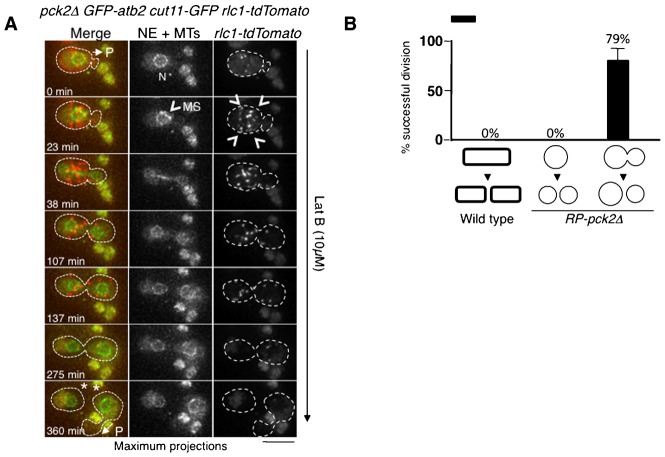
Fig. 5. Cell division in the absence of actomyosin ring in RP-pck2Δ cells.
(A) Time-lapse images of protruding pck2Δ cells expressing GFP-Atb2, Rlc1-tdTomato and Cut11-GFP as markers of the mitotic spindle, actomyosin ring and nuclear envelope, respectively. Cells were treated with 10 µM of LatB and recorded in multiple focal planes every 7.5 minutes. Maximum z-projections of representative time points are shown. P indicates the formation of a protrusion at time 0 and time 360 min, the asterisk denotes cell separation and N indicates the position of the nuclei. Arrowheads denote the myosin spots at the cell cortex indicative of an unassembled actomyosin ring. MS indicates the formation of the mitotic spindle at time 23 min. Dashed line highlights the cell border. Scale bar: 5 µm. (B) Percentage of pck2Δ and RP-pck2Δ cells undergoing successful divisions in the presence of 10 µM of LatB.

