
Figure 1.
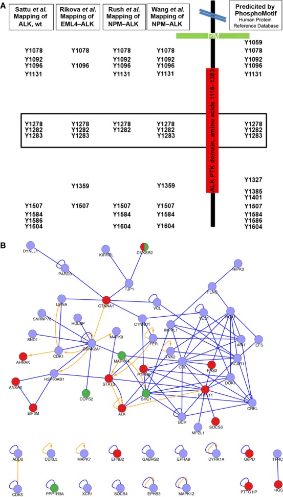
(A) Tyrosine residues phosphorylated in the kinase domain of ALK. The intracellular domain of ALK containing the protein kinase domain (PKD) (red) and potential autophosphorylation sites were searched with phosphomotif (http://www.hprd.org/PhosphoMotif_finder) as indicated. Presented and compared side-by-side with our phosphotyrosine mapping of activated full-length ALK are the global surveys of phosphotyrosine peptides identified in EML4–ALK and NPM–ALK 29,30. The critical tyrosines in the activation loop of the kinase domain of ALK are boxed 45. (B) Protein–protein interactions of human orthologs of the phosphoproteins identified in ALK-expressing PC12 cells. In the network, proteins with upregulated phosphorylation sites in activated ALK-expressing PC12 cells as compared with control cells are in red, and proteins with downregulated phosphorylation sites are in green. Blue edges indicate protein–protein interactions, and orange edges indicate kinase–substrate relationships. Only the network including ALK is shown. The pale blue balls indicate (human orthologs of) PC12 proteins with identified/mapped phosphotyrosines that were not found to be significantly regulated.