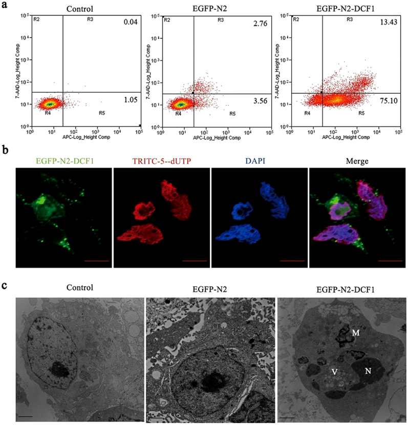
Figure 3. DCF1 caused U251 cell apoptosis.
(a). After transfection of dcf1 for 48 h, the early apoptosis accounting for about 75% and the late apoptosis accounting for about 13% were analyzed by Annexin V-APC/7-AAD flow cytometry detection. (b). Late cell apoptosis (red) was analyzed by TUNEL methods after transfection of dcf1 for 72 h. Scale bars,10000 nm (c). TEM revealed the occurrence of apoptosis, such as nucleus fragmentation(N), intracellular vacuolization(V), and autophagosome formation(A). Scale bars, 1 μm.

