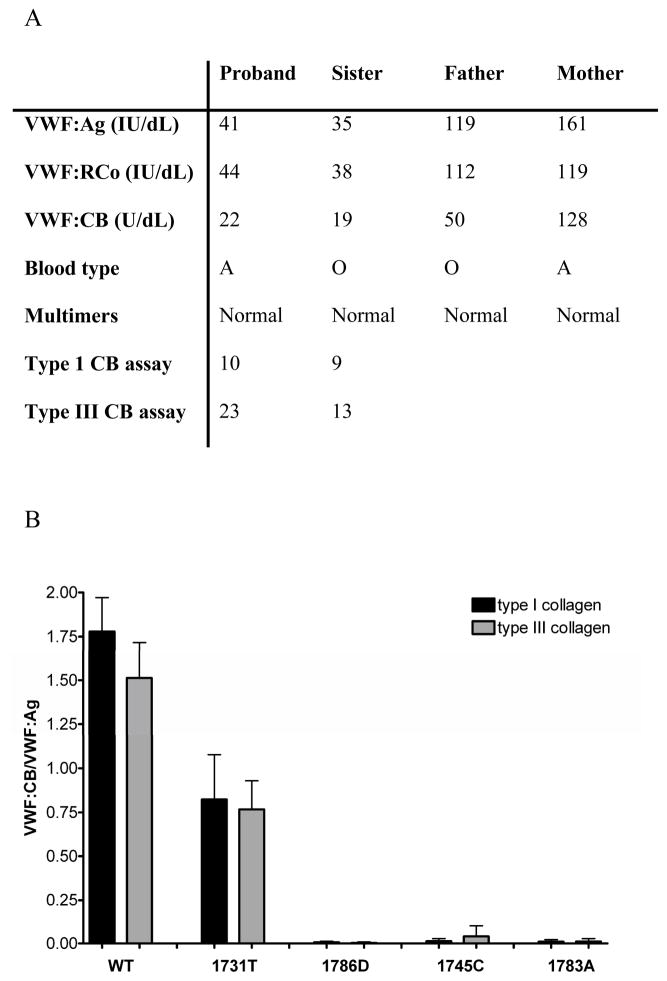
Figure 1.
Panel A shows VWF levels for the H1786D proband and family members. VWF:Ag, VWF:RCo, and VWF:CB listed for each subject were performed in the clinical laboratory along with blood type and multimer analysis. The VWF:CB used type III collagen. In addition, plasma samples from the proband and sibling were tested in the research laboratory using collagen binding assays for type I and type III collagen. Results are the mean of 3 separate assays. Panel B shows collagen binding for the recombinant VWF constructs. Binding to type I collagen is shown in black and binding to type III collagen is shown in grey for wild-type (WT), 1731T, 1786D, 1745C, and 1783A VWF. Error bars represent 1 SD for a minimum of 3 separate assays.

