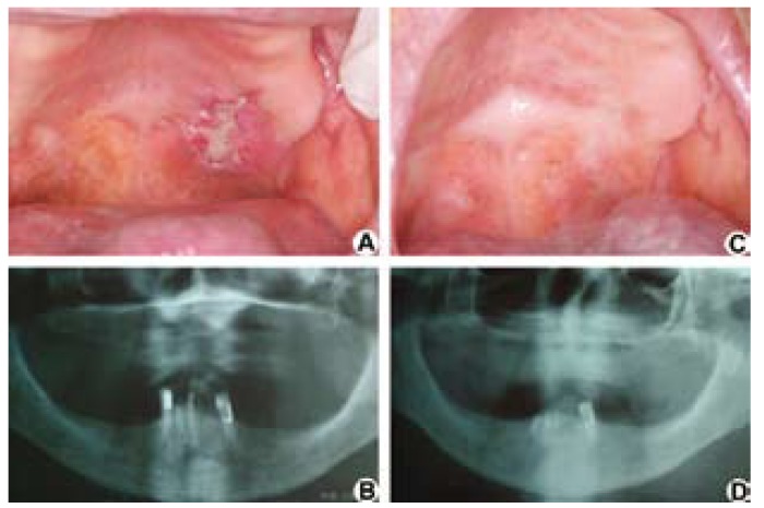
Figure 1.
A) Initial clinical image. An ulcer of raised and irregular edges could be observed on the border of the hard and soft palates. B) Initial panoramic radiographshowed no bone changes. C) Clinical image and D) panoramic radiograph after thirty-one months of diagnosis and treatment, in which the lesion’s total clinical resolution could be observed.

