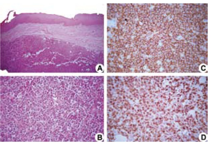
Figure 2.
Histopathological sections stained with hematoxylin-eosin show a neoplastic proliferation of large lymphoid cells (A, 50x original magnification) and the presence of neoplastic cells with coarse chromatin and inconspicuous nucleoli (B, 400x original magnification). Immunopositive cells of anti-CD20 (C, 400x original magnification, Streptoavidin-biotin) and anti-Ki-67 (D, 400x original magnification, Streptoavidin-biotin).

