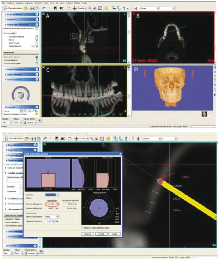
Figure 1.
The computer program employed in the present study (SimPlant® - Materialise-Leuven-Belgium) allows visualization of inter-radicular spaces in a multitude of 2-D and 3-D points of view. The cross-sectional (A), axial,(B), panoramic (C) and 3-D (D) images are visible at the same time on a computer monitor. Lower image: The cross-section image from SimPlant used to measure the cortical bone thickness at 2, 4, 6 and 8 mm from the alveolar crest.

