
Figure 7.
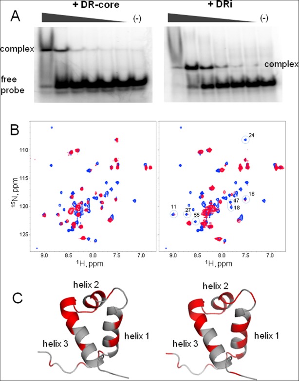
Binding of the PAI subdomain of the DNA-binding domain of SB transposase to DR-core (left panel) and DRi (right panel) DNA sequences. A: EMSA shows that the PAI subdomain binds to the DR-core and DRi DNA. Protein–DNA complexes were formed in 20 mM HEPES buffer at pH 7.5 in the presence of 0.1 mg/mL BSA, 1 µg [di:dC], 1 mM DTT, 150 mM NaCl, and 1 mM MgCl2. Probe quantity was fixed at 0.21 pM and the quantities of PAI subdomain varied from 0 to 2.24 nM as indicated by triangles above gels. B: [15N,1H]-HSQC spectra of pure PAI subdomain (blue) and of the PAI subdomain in the presence of a twofold molar excess of DR-core and DRi DNA sequences. The spectra were collected at 5°C in aqueous (5% D2O/95% H2O) 25 mM sodium phosphate buffer at pH 7.0 in the presence of 300 mM NaCl. C: Cartoon structures of PAI subdomain show residues affected by the binding of DR-core (left) and DRi (right) DNA sequences in red.