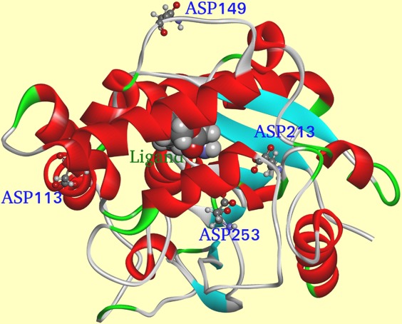
Figure 3.

Cartoon representation of LipK107 structure. Residues selected by our ETSS software were shown in stick model and labeled. The ligand is shown in ball model and labeled. α-Helices are colored in red, β-strands are colored in blue, and random coils are colored in green.
