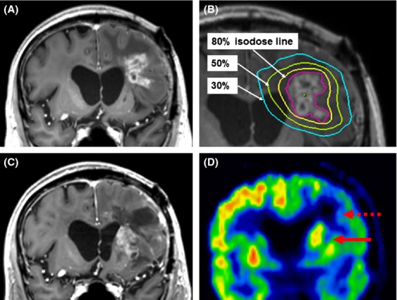
Figure 3.

Representative case of marginal recurrence. (A) A recurrent tumor from an anaplastic oligoastrocytoma located in the left frontal lobe, adjacent to the initial surgical cavity and within the field of the initial radiotherapy. (B) The tumor was treated with salvage stereotactic radiotherapy (SRT). Target delineation of the planning target volume (PTV, indicated by the magenta line) was classified as method A (contrast-enhancing lesion plus a 1-mm margin); the prescribed dose was 35 Gy in five fractions with 80% coverage of the PTV. (C) At 10 months after treatment, a new recurrent tumor at the left basal ganglion emerged, adjacent to the previously treated lesion (marginal recurrence). (D) L-Methyl-11C-methionine positron emission tomography supported the diagnosis of recurrence (indicated by a solid arrow), while the SRT-treated area was determined to be radiation necrosis (indicated by a dashed arrow).
