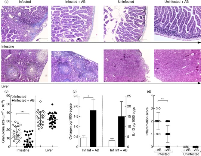
Fig. 5.
Infected antibody-treated mice present reduced intestinal inflammation and granuloma formation but elevated collagen levels in hepatic granulomas. (a) Histological sections were prepared from the intestines (upper panel) or liver (lower panel) from control and 8-week-infected mice and stained with Masson's Blue. Magnification was ×40 of equally sized sections. Organ sections from infected mice are depicted far left; infected and antibiotic treatment groups, left; naive mice, right; and naive mice treated with antibiotics, far right. (b) From four independent infection studies, the average size of 35 liver granulomas and up to 15 intestinal granulomas were measured in each infected mouse. Symbols represent mean ± standard deviation (s.d.) of individual mice (–AB n = 22 and +AB n = 28). (c) The amount of collagen was determined in individual liver samples using the Biocolor assay kit and then correlated to the number of eggs found in the livers of the corresponding mice. The results shown are the mean ± s.d. obtained from individual mice from two independent infection experiments. In-situ levels of interleukin (IL)-13 were measured in the supernatants of liver homogenates at the time of analysis by enzyme-linked immunosorbent assay (ELISA). Bars represent the mean ± standard deviation (s.d.) of individual mice from three independent infection studies. (d) Semi-quantitative pathological assessment of intestinal inflammation. In a blind fashion, intestinal sections from individual mice were assessed for their level of inflammation (0 = absent; 1 = mild, 2 = moderate inflammation and 3 = strong inflammation). Symbols represent mean ± standard deviation (s.d.) of Inf. n = 10; Inf + AB n = 15; naive n = 5 and naive + AB n = 5). Asterisks indicate a significant difference (*P < 0·05; **P < 0·01; ***P < 0·001) between the indicated groups following analysis of variance (anova) or t-test analysis.

