Fig. 5.
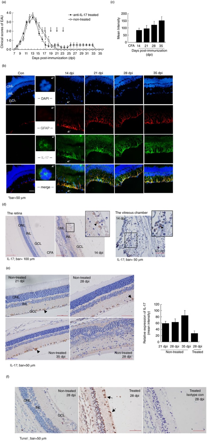
Neuroprotection effect of interleukin (IL)-17 in the late phase of an acute experimental autoimmune uveitis (EAU) model. (a) Clinical scores of the EAU in Lewis rats with or without the treatment of anti-IL-17 antibodies. The animals in the treatment group received intravenous injections of anti-IL-17-neutralizing antibody (20 μg/rat) on the 17, 19, 21 and 23 days post-immunization (dpi); the non-treated rats received phosphate-buffered saline (PBS). The mean score at each dpi is the average for five to seven animals. (b) Immunofluorescent microscopy with anti-glial fibrillary acidic protein (GFAP) (red) and anti-IL-17 antibodies (green) showed a significant number of double-positive staining cells in the EAU versus control retina from 14 to 35 dpi, indicating the expression of IL-17 in the reactive astrocytes during the development of disease. At 14 dpi, a significant single-positive staining of IL-17 on the infiltrating cells was noticed (2nd column, arrows). (c) The double-positive staining astrocytes were quantified by measuring the yellow (merged with green and red) fluorescent intensity in the total retinal area using NIS-Elements BR version 4·00·00 software [means ± standard error of the mean (s.e.m.), n = 10]; *P < 0·05. (d) Immunohistochemistry with an anti-IL-17 antibody shown at 14 dpi, the peak of disease. Intensive IL-17-positive staining was observed in the infiltrating immune cells (in the retina and the vitreous chamber) and resident neural cells (the arrows). (e) Immunohistochemistry staining with an anti-IL-17 antibody at the resolution phase of EAU with or without treatment by neutralizing anti-IL-17 antibodies. The cytoplasm staining was evident at the GCL of the neural cells (the arrows). (f) Significant neural cell apoptosis in EAU treated with an anti-IL-17-neutralizing antibody (20 μg/per animal) by TUNEL (terminal deoxynucleotidyl transferase (TdT)-mediated dUTP nick end-labelling) assays. GCL: ganglion cell layer; INL; inner nuclear layer; ONL: outer nuclear layer. Data shown were means ± s.e.m. of at least three experiments.

