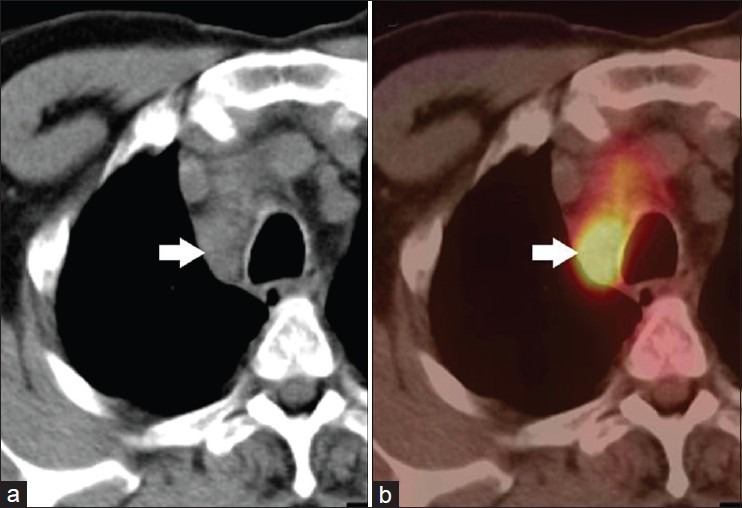
Figure 2.

A 52-year-old male with NSCLC of right lung. FDG PET-CT was done for staging. CT (a) showed enlarged right paratracheal node (arrow) which was FDG avid (SUVmax-9.2) on PET-CT (b). A diagnosis of nodal metastasis was made on PET-CT. However, this turned out to be tuberculosis at histopathology. Hence, results of FDG PET-CT for nodal staging should be confirmed with FNAC/biopsy to avoid false positives
