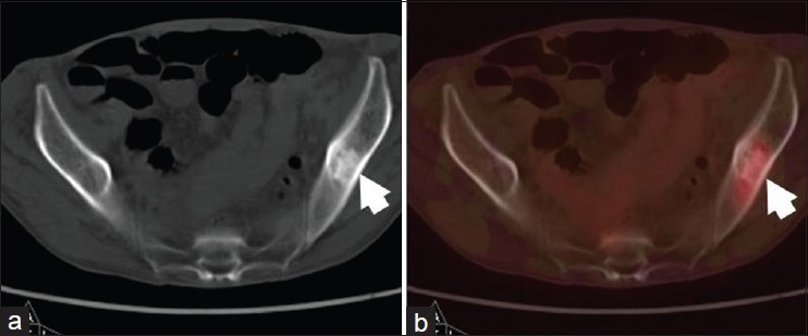
Figure 4.

A 70-year-old male with adenocarcinoma of right lung, postpneumonectomy and adjuvant radiotherapy. He presented with bony pains. FDG PET-CT was done to rule out distant metastasis. On CT (a) images a sclerotic lesion was seen in left ilium (arrow). It showed mild FDG uptake on PET-CT (b) images, suggesting skeletal metastasis
