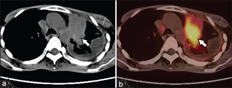
Figure 5.

A 49-year-old male, postsurgery and radiotherapy for left lung NSCLC. PET-CT was done 9 months later for restaging. CT (a) images showed mass lesion in the thorax with fibrotic changes in pleura. PET-CT (b) images showed intense FDG uptake (SUVmax-13) in the mass suggesting recurrent disease (arrow). No uptake was noted in the pleura suggesting post therapy changes
