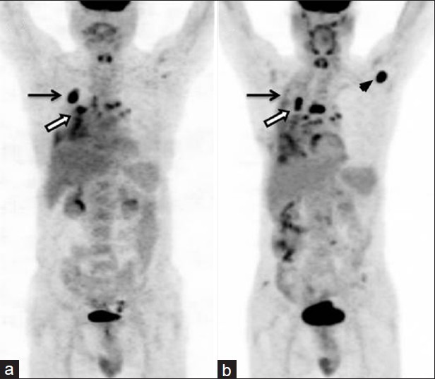
Figure 6.

A 51-year-old male with right lung NSCLC with nodal metastasis. FDG PET-CT (a) showed primary lung lesions (arrow) with mediastinal nodal metastasis (bold arrow). He underwent three cycles of chemotherapy. Post therapy PET-CT (b) showed almost complete regression of primary lesion (arrow) but increase in size and uptake of mediastinal nodes (bold arrow). Also noted was appearance of new axillary nodal metastasis (arrowhead), suggesting progression of disease
