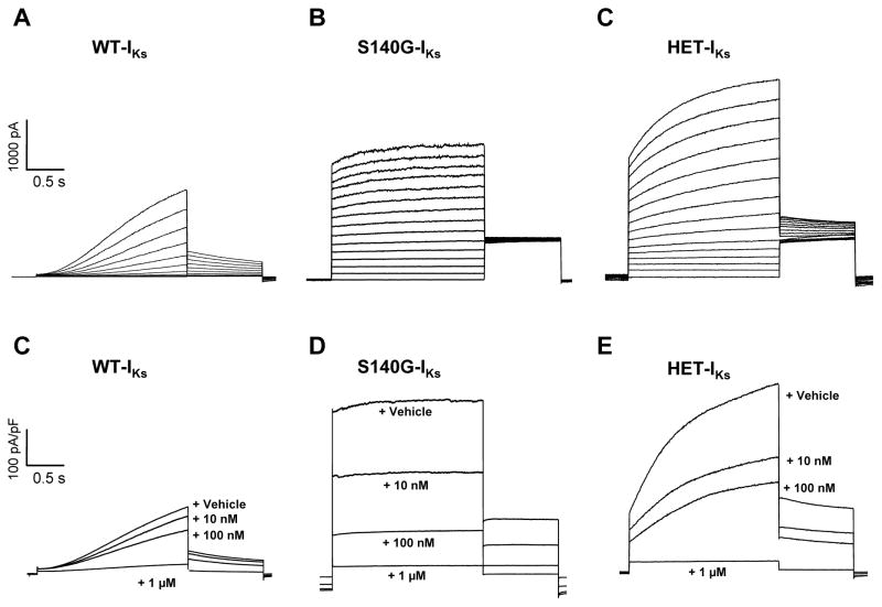
Figure 1.
S140G-IKs and HET-IKs exhibit enhanced sensitivity to HMR-1556. A, B and C, Representative current recordings from cells expressing WT-IKs (A), S140G-IKs (B), or HET-IKs (C). Recordings illustrated in A, B and C were obtained using the activation protocol described in the Methods. D, E, and F, Average current densities (current normalized to cell capacitance) elicited by a 2 s voltage step to +40 mV followed by a 10 s interpulse during application of vehicle or various concentration of HMR-1556 from cells expressing WT-IKs (D), S140G-IKs (E), or HET-IKs (F). Current density traces in D, E, and F are averages from 9–11 cells.

