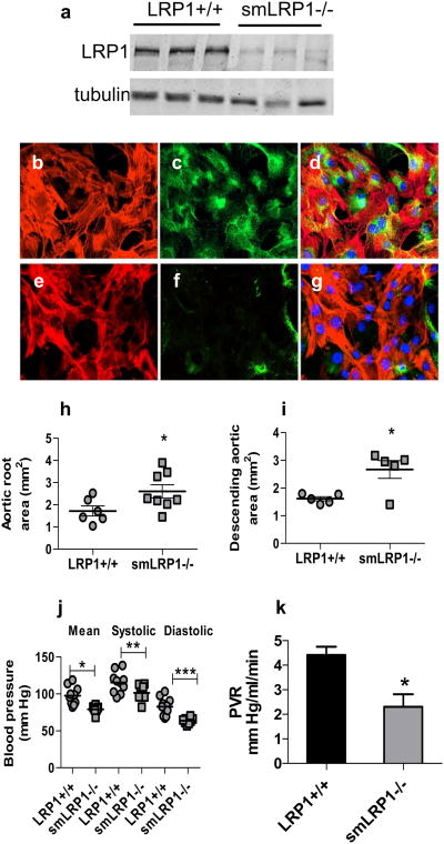
Figure 1. SMC LRP1 deficiency induces aortic dilatation.
a) Immunoblot analysis of aortas (adventitia removed) from LRP1+/+ and smLRP1-/- mice. a/b tubulin was used as a loading control. b–g) Immunofluorescent analysis of SMC isolated from aortas of LRP1+/+ (b, c, d) and smLRP1-/- (e, f, g) mice. Cells were stained for αSMA (red, b, e) and LRP1 (green, c, f). Merged images are shown in d and g. h) Aortic root area (*p=0.04) and i) thoracic aortic area (*p=0.01) as measured by echocardiography in 20 weeks old mice; j) blood pressure measurements were performed by cannulating the right carotid artery and recording the blood pressure continuously (*p=0.001, **p=0.03, ***p=0.0006); k) peripheral vascular resistance for LRP1+/+ and smLRP1-/- mice. *p=0.014, n=4)

