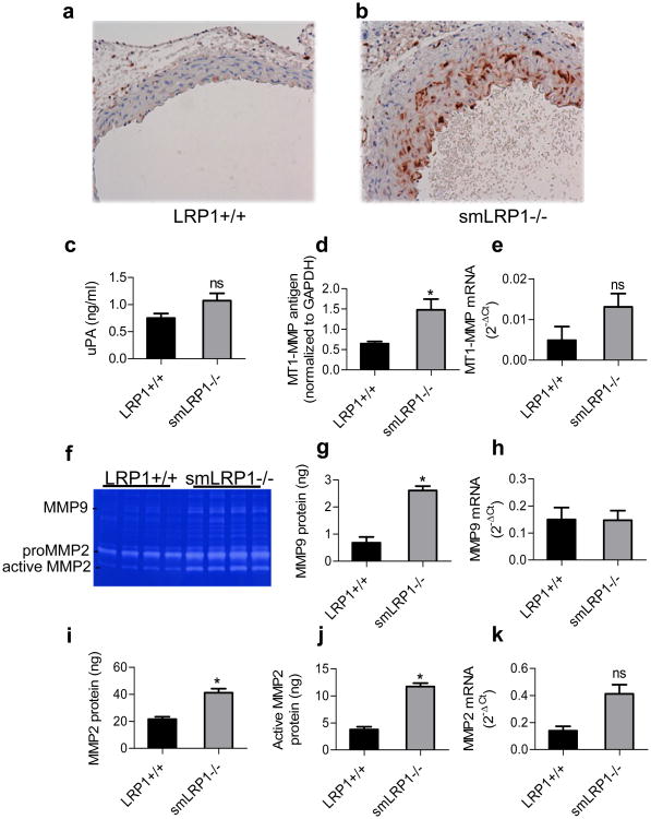
Figure 5. Increased macrophage infiltration and proteinases expression in aortas from smLRP1-/- mice.
Abundant Mac-2 expression was detected in smLRP1-/- aortic vessel wall (b) while none was detected in WT (a) aortas. c) Expressions of uPA determined by ELISA; d) MT1-MMP levels were quantified by immunoblot analysis of aortic extracts; e) MT1-MMP mRNA levels determined by qRT-PCR analysis; f) Gelatin zymography analysis of aortic extracts reveal increased levels of MMP9, pro-MMP2, and active MMP2; g,i,j) levels of MMP9, MMP2 and active MMP2 were quantified by gelatin zymography using purified proteins as standards. (g, *p=0.0004, n=4; i, *p=0.001, n=4; j, *p<0.0001, n=4); h) mmp9 mRNA levels determined by qRT-PCR analysis; (*p=0.002); k) mmp2 mRNA levels determined by qRT-PCR analysis (FDR adjusted p=0.08).

