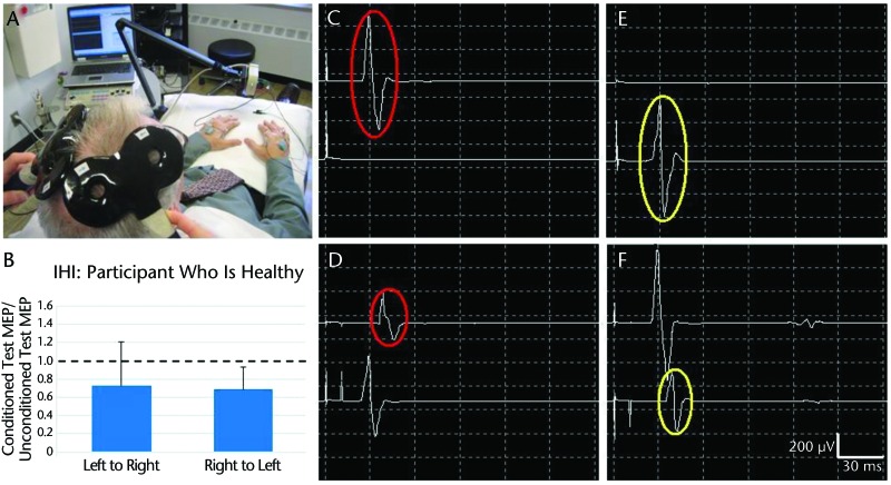
Figure 1.
Interhemispheric intervention (IHI): participant who is healthy. (A) Setup for measuring IHI with two 50-mm figure-8 coils over left and right primary motor areas (M1) of participant who is healthy and electromyography electrodes on bilateral first dorsal interosseous (FDI) muscles. (B) Mean (SD) values showing relatively balanced IHI in both directions, derived from multiple trials of left M1 inhibiting right M1 (left to right) (C–D) and right M1 inhibiting left M1 (right to left) (E–F). (C) Motor-evoked potential (MEP) in left FDI (upper trace) in response to unconditioned suprathreshold test stimulus to right M1. (D) MEP in right FDI (lower trace) in response to suprathreshold conditioning stimulus to left M1, followed by a MEP in left FDI in response to test stimulus to right M1 (interstimulus interval=10 ms). Interhemispheric intervention in the direction of left M1 inhibiting right M1 is demonstrated by reduction in conditioned test MEP (lower red circle) compared with unconditioned test MEP (upper red circle). (E–F) Interhemispheric intervention in the direction of right M1 to left M1 (compare yellow circles).

