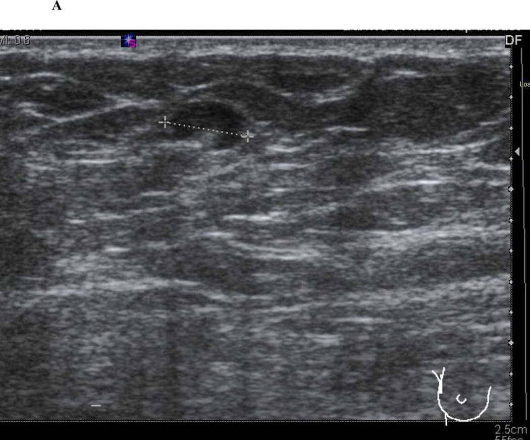
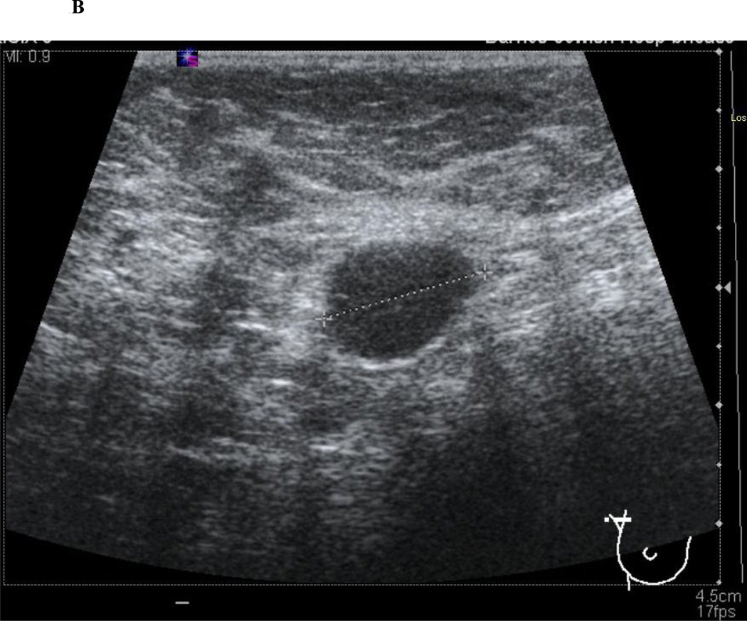
Figure 1.
Axillary ultrasound characteristics of normal and abnormal lymph nodes. Normal lymph nodes have a smooth, homogenous cortex with a centrally located, preserved fatty hilum (1A). Abnormal, or suspicious for metastatic involvement, lymph nodes have a rounded appearance with an eccentrically thickened, heterogenous cortex and effacement of the fatty hilum (1B).


