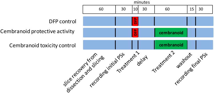
Figure 2.
The experimental protocol. The figure represents the three lanes of the incubation chamber; blue color stands for periods where the slice was superfused with ACSF. There were three treatment groups: 1) DFP control, where slices were exposed to 100 μM DFP for 10 min and superfused with ACSF for 105 min before recording the final PSs; 2) Cembranoid protective activity group, where slices were exposed to 100 μM DFP for 10 min, washed for 30 min and then exposed to 10 μM cembranoid for 1 h, and 3) Cembranoid toxicity control, where slices were exposed to 10 μM cembranoid for 1h.

