Fig. 1.

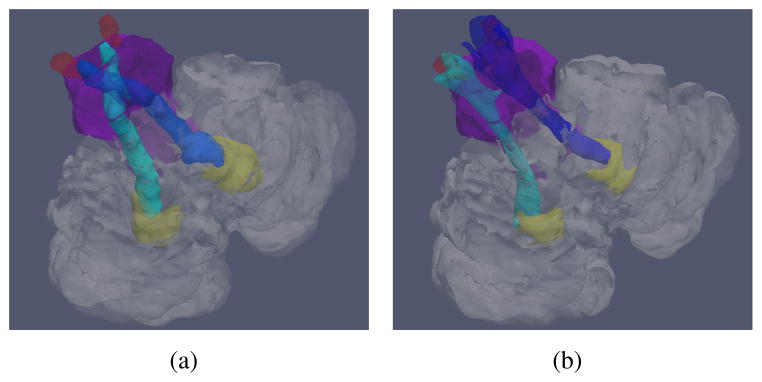
Illustrations of (a) the correct structures of the SCPs, and (b) typical incorrect SCPs obtained from DTI. Shown together with the cerebellum (transparent), the brainstem (purple), the DN (yellow), and the RN (red).

Illustrations of (a) the correct structures of the SCPs, and (b) typical incorrect SCPs obtained from DTI. Shown together with the cerebellum (transparent), the brainstem (purple), the DN (yellow), and the RN (red).