
Figure 1.
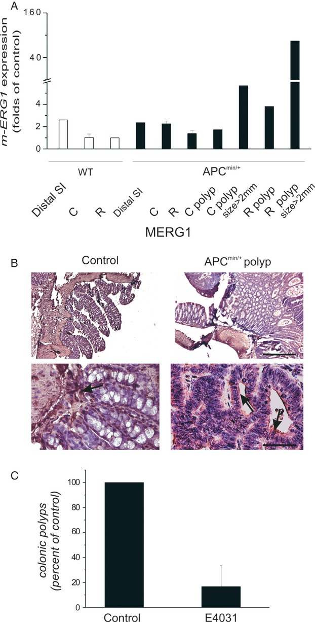
Expression and role of mERG1 in Apcmin/+ mice. (A) Analysis of mERG1 expression by RT-PCR in small and large intestine of WT and Apcmin/+ mice and in colonic and rectal polyps developed in Apcmin/+ mice after a normalization for mouse myh11, characteristic of myofibroblasts and smooth muscle cells, to detect the only mERG1 epithelial expression27. Distal SI, distal small intestine; C, colon; R, rectum; C polyp, colonic polyp; R polyp, rectal polyp. Data relative to colon and rectum derived from two different experiments, each carried out in triplicate, are reported as the mean ± SEM and were calibrated to the expression levels determined in the rectum of WT mice. Data relative to distal small intestine and polyps derived from a single experiment, carried out in triplicate, are reported as the mean, and were calibrated to the expression levels determined in the rectum of WT mice. (B) mERG1 expression in control (WT) and Apcmin/+ polyps was evaluated by IHC. An anti-hERG1 monoclonal antibody was used as detailed in Material and Methods. Upper panels: 50× magnification, bar: 200 μm; lower panels: 400× magnification, bar: 20 μm. (C) The number of colonic polyps obtained after E4031 treatment of Apcmin/+ mice. Four 1-month-old Apcmin/+ mice received daily IP injections of E-4031 for 3 months, while two Apcmin/+ mice received buffered saline only. After death, the number of polyps that developed in colon of Apcmin/+ mice was determined under a dissecting microscope (20× power field). Data were expressed as mean ± SEM.