
Fig. 6.
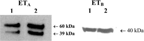
Identification of ETA and ETB receptor protein by Western blot using ETA (left) and ETB (right) receptor-specific antibodies. Lanes 1 and 2 represent myoFbs and coronary endothelial cells, respectively. ETA immunoblot with anti-ETA-specific antibody resulted in two bands of ≈60- and 39-kDa proteins, whereas immunoblot with anti-ETB receptor antibody identified a protein band of ≈40 kDa. Preimmune serum served as a control (not shown).