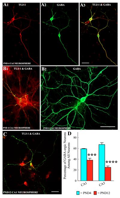
Figure 4.
Neurospheres derived from CA1 and CA3 subfield cells of postnatal day 4 (PND4) and PND12 hippocampi generate GABA-ergic interneurons. Figures A1-A3 show examples and morphology of TuJ-1 positive neurons generated from PND4 CA3 neurosphere cells that expressed GABA. Figure B1 illustrates GABA-ergic axon terminals on soma and dendrites of non-GABA-ergic hippocampal pyramidal-like neurons derived from PND4 CA1 neurosphere cells. B2 is an example of a well-developed GABA-ergic neuron generated from a PND4 CA3 neurosphere cell. Figure C shows an example of a well-developed GABA-ergic neuron (orange) generated from a PND12 CA1 neurosphere cell. Scale bar = 50μm. The bar chart in D shows percentages of GABA-ergic interneurons (among TuJ1+ neurons) derived from neurospheres of CA1 and CA3 subfield cells of PND4 and PND12 hippocampi. Note decreased percentages of GABA-ergic neurons in neurospheres of PND 12 subfields (25-38%), in comparison to neurospheres of PND 4 subfields (59-67%). ***, p<0.001; ****, p<0.0001.

