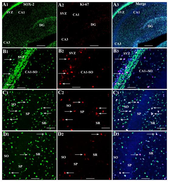
Figure 7.
Proliferating NSC-like cells in the postnatal day 4 hippocampus visualized through Sox-2 and Ki-67 dual immunofluorescence and Z-sectioning analyses in a confocal microscope. Figures A1-A3 illustrate Sox-2+ cells and Ki-67+ cells in hippocampal regions as well as the posterior subventricular zone (SVZ) overlying the CA1 region. Figures B1-D3 show cells expressing both Sox-2 and Ki-67 in the posterior SVZ (arrows in B1-B3), strata oriens (SO) radiatum (SR) of CA1 subfield (arrows in C1-C3), and the SR and stratum pyramidale (SP) of the CA3 subfield (arrows in D1-D3). Scale bar, A1-A3 = 100μm. B1-D3 = 50μm.

