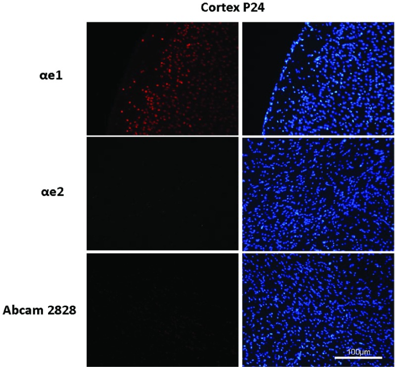
Figure S4. Staining of the cortex of a P24 WT mouse with isoform-specific antibodies.
20 μm brain sections from a C57 male MeCP2 WT mouse were prepared as described in materials and methods. These sections were stained with αe1 (top section), αe2 (middle section) and Abcam2828 (bottom section), followed by anti-rabbit Alexa 546 secondary antibody, and mounted in DAPI-containing mounting medium.

