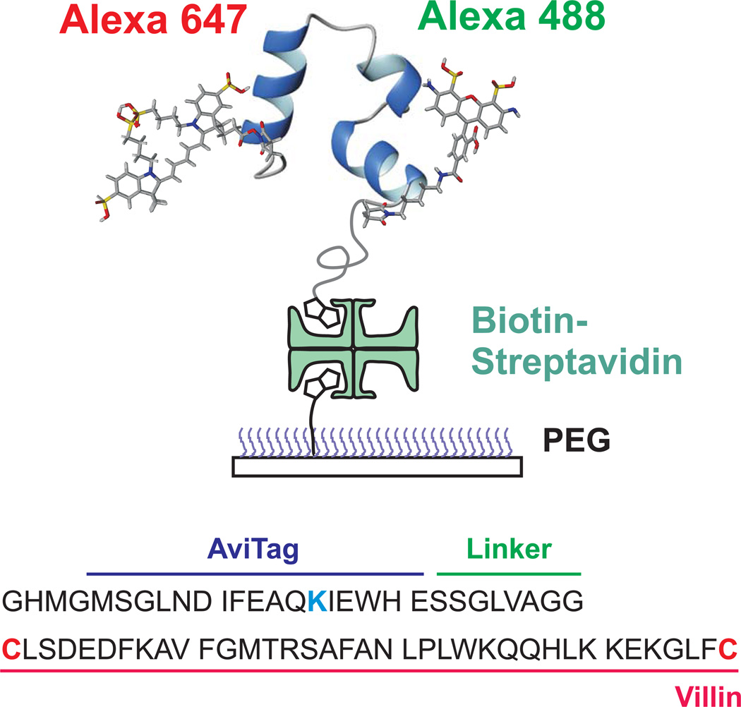
Fig. 1.
Schematic of immobilized folded villin subdomain showing donor (green-emitting) and acceptor (red-emitting) fluorophores. Protein molecules were attached to a polyethyleneglycol-coated glass surface via a biotin-strepavidin-biotin linkage. Dyes were attached to cysteine residues (red) at the N- and C-termini of villin and a biotin molecule to the lysine residue (blue) within the AviTag sequence.

