
Figure 1.
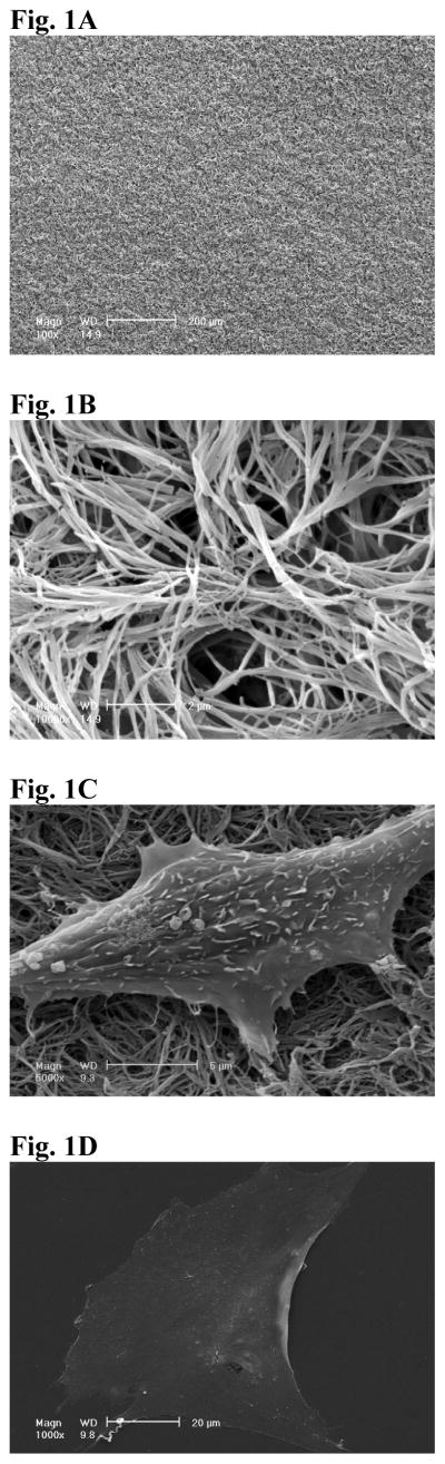
SEM view of NF matrix at low magnification (A) and high magnification (B). The average diameter of the fibers of PLLA NF matrix was between 100 and 200 nm. SEM view of MC-4 cells cultured on NF matrix (C) and flat films (D) for 24hr. The cells on NF matrix were more rounded, with small processes interacting with PLLA nanofibers.