
Fig. 3.
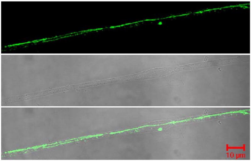
ZO-1 localization within the microvessel. Confocal micrographs reflect ZO-1 protein localization as continuous and junctional within the TJ. Phase contrast micrographs show the microvessel purity, and the merged micrograph demonstrates ZO-1 expression only within the microvessel. Micrographs were taken with an LSM 510 confocal laser scanning microscope at 63× with an oil immersion lens.