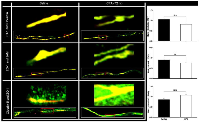
Fig. 6.
Co-localization of ZO-1 with transmembrane TJ proteins. Confocal micrographs demonstrate co-localization of ZO-1 with each transmembrane protein – occludin, JAM-1 and claudin-5 – in isolated microvessels from both saline and CFA (72 h) treated rats. The representative micrographs, taken at 100× with an oil immersion lens on an LSM 510 confocal laser scanning microscope, qualitatively confirm the changes in protein expression seen by western blot for transmembrane TJ proteins. Higher magnification micrographs correlate to portions of the inset microvessels, outlined in red. A 1:1 ratio of protein co-localization with ZO-1 appears yellow, and any alterations from there are evident as increased green or red fluorescent channel. MetaMorph software was used to examine the relative fluorescence intensities of each channel, and is expressed as ratios in terms of the proteins they represent. These ratios demonstrate decreased co-localization of ZO-1 with both occludin and JAM-1, and an increase with claudin-5. Confocal micrographs of cerebral capillaries (vessel diameter 4–10 μm) are representative of at least 3 separate and independent experiments; n≥6 per group, *p<0.01, **p<0.001. (For interpretation of the references to colour in this figure legend, the reader is referred to the web version of this article.)

