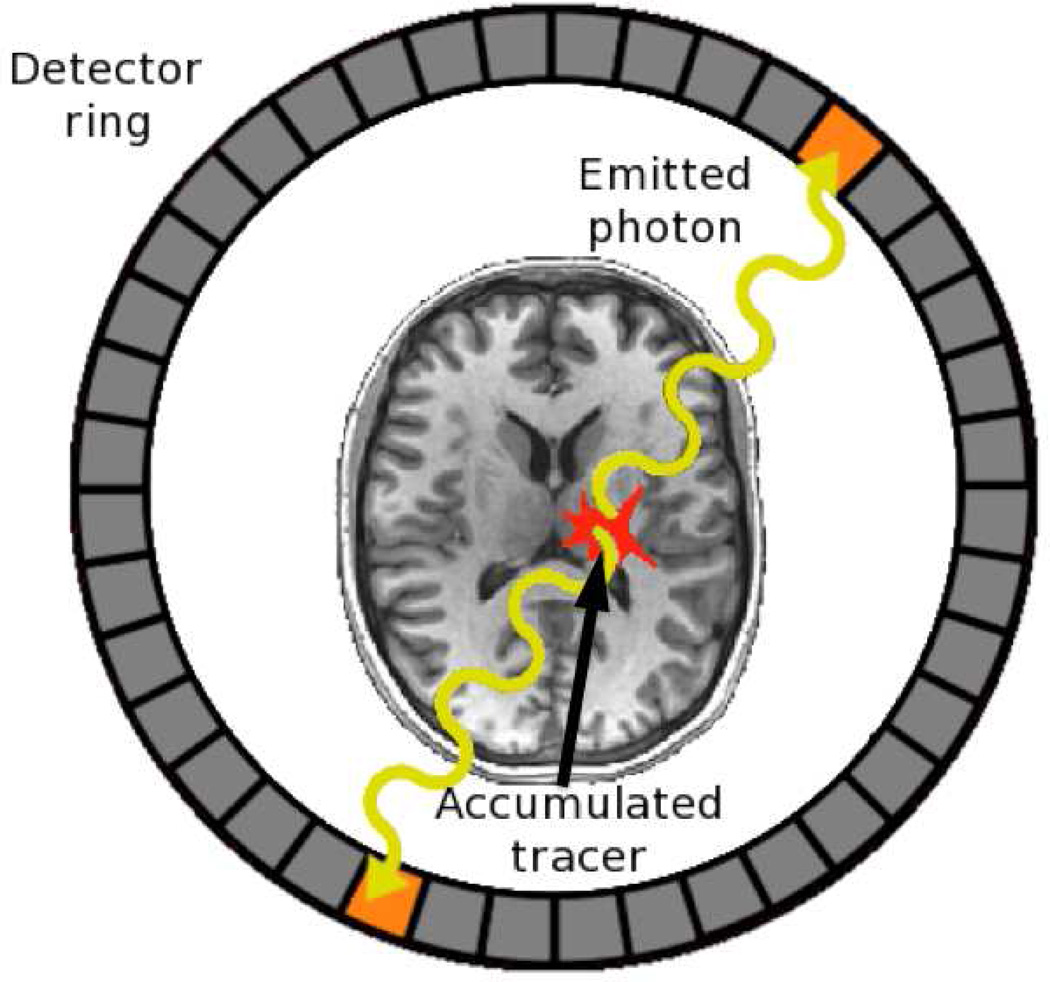
Figure 1.
Coincidence detection in positron emission tomography (PET). Accumulated radioactive tracer within a patient releases positrons that annihilate with nearby electrons, resulting in the emission of two high-energy photons (gamma rays) that intersect with a detector ring surrounding the patient, with the detections being coincident in time. The two detectors form a line along which the positron annihilation must have occurred. The intersection of many such detected coincidence lines, observed over the course of a scan, serves to pinpoint areas of relatively high concentration of the radioactive label. Two- or three-dimensional images indicating where the labeled compound (e.g., 15O-labeled water) accumulated in the brain may then constructed by back-projection.

