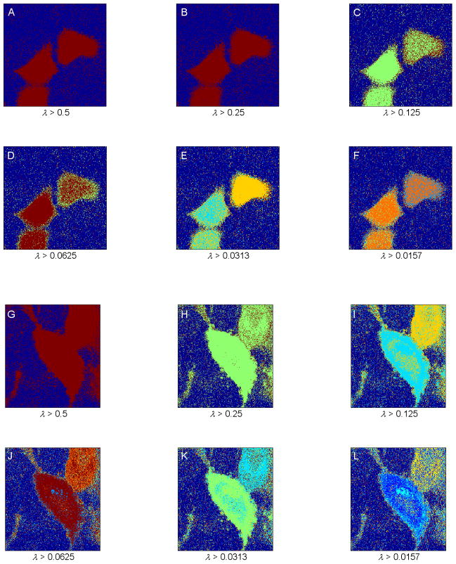
Fig. 4.
(A–F) Segmentation of the fluorescence lifetime imaging microscopy (FLIM) image shown in Fig. 1A, using the spectral clustering method developed by Ng et al., 2002 for increasing resolution. False colors represent different segments. Spectral clustering was not able to provide the expected segments, and it introduced a high amount of noise in the segmented images. (G–L) Segmentation of the FLIM image shown in Fig. 1B, using the same spectral segmentation method for increasing resolution. False colors represent different segments. The segmented images show the two cells in a single segment in low resolution (G–H) and in two distinct segments in a slightly higher resolution (I–J). At the resolution limit, similar to the case for the image shown in Fig. 1A, the segmented images display a high amount of introduced noise.

