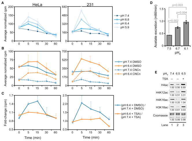
Figure 6. Efflux of Acetate through the MCTs at Low pH Is Dependent on HDAC Activity.
(A–C) Liquid scintillation counts (mean ± SD) of media from HeLa and 231 cells labeled with 3H-acetate and treated as indicated in 10% FBS-containing DMEM.
(A) Cells were exposed to media of different pHe for the indicated time. (B) Cells were treated with the MCT inhibitor CNCn (10 mM) or dimethyl sulfoxide during the chase and during subsequent treatment at the indicated pHe. (C) Cells were treated with 500 nM TSA or DMSO prior to labeling and during subsequent treatment at varying pHe.
(D) Acetate excretion rate (mean ± SD) in 231 cells incubated for 30 min in 10% FBS-containing DMEM without P at the indicated pHe.
(E) WBs of histone acetylation in 231 cells treated for 16 hr in complete DMEM at the indicated pHe followed by treatment at the same pHe with or without 500 nM TSA. See also Figure S5.

