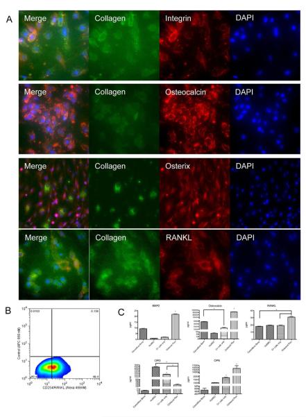
Figure 5. Proteomic profiling of Osteocel Plus cells cultured for 28 days in ODM.
(A) Immunofluorescent staining (20x) for the osteoblast markers Collagen (Alexa 488), Integrin (Cy3), Osteocalcin (Cy3), Osterix (Cy3), RANKL (Cy3), and for nuclear DAPI (blue). (B) Flow cytometry analysis for CD254/RANKL. (C) ELISA and multiplex immunoassay for BMP-2, Osteocalcin, RANKL, Osteoprotegrin (OPG), and Osteopontin (OPN). Comparative analysis was performed on acellular cancellous bone, huMSCs, Osteocel Plus cells only, and native Osteocel Plus cells adherent to the bone. Concentrations are expressed as pg/ml. (n=3, *p<0.05, 95% Confidence interval of difference).

