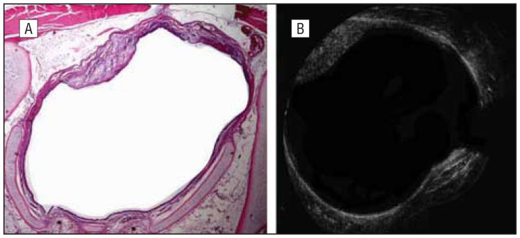
Figure 7.
A rabbit killed 42 days after mucosal injury. Histologic (A) and optical coherence tomographic (OCT) (B) images show a single area of fibrous scarring at the 11-o'clock position. The OCT image very clearly demonstrates changes in both surface and submucosal anatomy that correlates well with histologic images.

