Fig. 10.
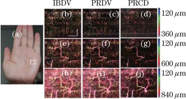
(Color online) En-face MIP view image of human palm skin. (a) Photograph of the imaging area (in the white rectangle). En-face MIP view of IF-IBDV images for depths of (b) 120–360, (e) 120–600, and (h) 120–840 μm; En-face MIP view of IF-PRDV images for depths of (c) 120–360, (f) 120–600, and (i) 120–840 μm; En-face MIP view of IF-PR-D-OCT images for depths of (d) 120–360, (g) 120–600, and (j) 120–840 μm[45]. Scale bar: 1 mm.

